Live-Cell-Imaging Core Lab
Committee:
- Dr. Yu, Tien-Shin(chair)
- Dr. Chiou, Jian-Geng
- Dr. Jauh, Guang-Yuh
- Dr. Verslues, Paul E.
- Dr. Wang, Chung-Ju
Research Assistant
Live-cell imaging has become one of the most important techniques in modern cell biology. The Live-Cell-Imaging Core Laboratory of the Institute of Plant and Microbial Biology provides users with on-site tutorial services and technical supports on light and fluorescence microscopy, Delta Vision microscopy, confocal microscopy and laser microdissection and capturing technology.
Facilities:
-
Confocal Laser Scanning Microscope::
- 1. Olympus FV3000 (Invert 2022) lasers: 405 nm, 445 nm, 488 nm, 514 nm, 561 nm, 594 nm and 640nm
- 2. LEICA STELLARIS 8 (Invert 2020) Diode 405nm WHITE LIGHT LASER 440-790nm
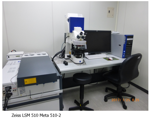
- 3. Zeiss LSM 510 Meta (upright, 2007), Diode 405nm; Argon 458/477/488/514 nm; HeNe/543 nm; HeNe/633 nm
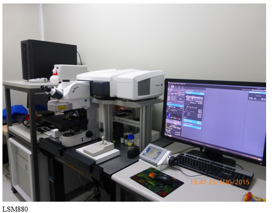
- 4 .Zeiss LSM880 with Airyscan (upright, 2015) , Diode 405nm; Argon 458//488/514 nm; DPSS 561 nm; HeNe/633 nm
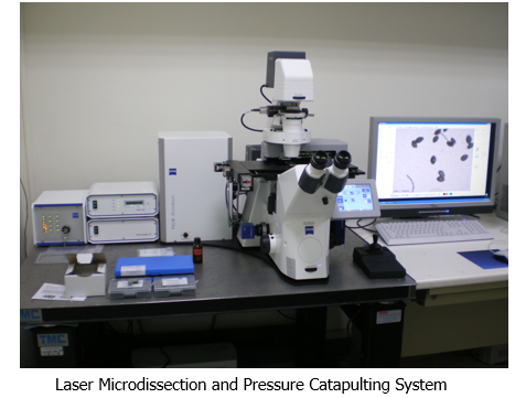
- Laser Microdissection and Pressure Catapulting System (Zeiss Palm, 2008)
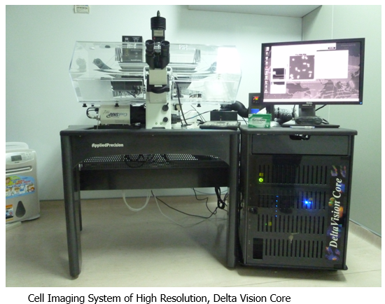
- Cell Imaging System of High Resolution, Delta Vision Core (Applied Precision Inc. Delta Vision Core, 2010)
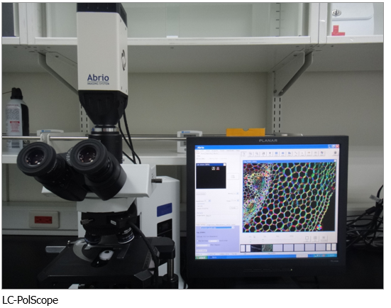
- Polarizing Microscope and Abrio Imaging System(LC-PolScope, 2007)
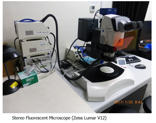
- Stereo Fluorescent Microscope (Zeiss Lumar V12, 2005)
- Inverted Fluorescent Microscope (Zeiss Axiovert S100, 1999)
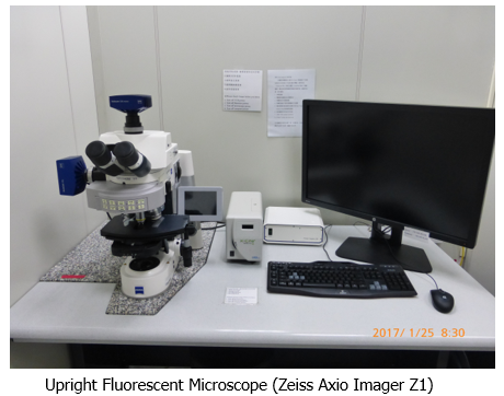
- Upright Fluorescent Microscope (Zeiss Axio Imager Z1, 2005)
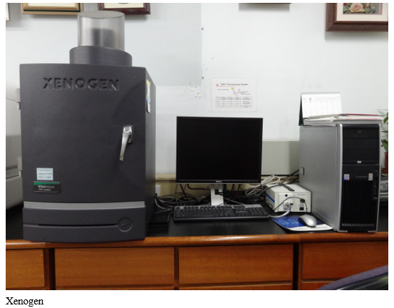
- Quantitative Fluorescence and Bioluminescence Imaging(XENOGEN IVIS System, 2006)
- Workstation with MetaMorph & Imaris image analysis.
Live-Cell-Imaging Core Lab (IPMB)
Operation manuals::
IPMB_FV3000_基本操作手冊_compressed.pdf
LEICA STELLARIS 8 (Invert, 2020), Diode 405nm; WHITE LIGHT LASER 440-790nm
ZEISS LSM 880
LSM880
Fluorescence probes for live cell imaging
GE DeltaVision DeltaVision 2019
LSM 510 META
Upright ZEISS AxioImager Z1
Laser Microdissection and Pressure Catapulting System
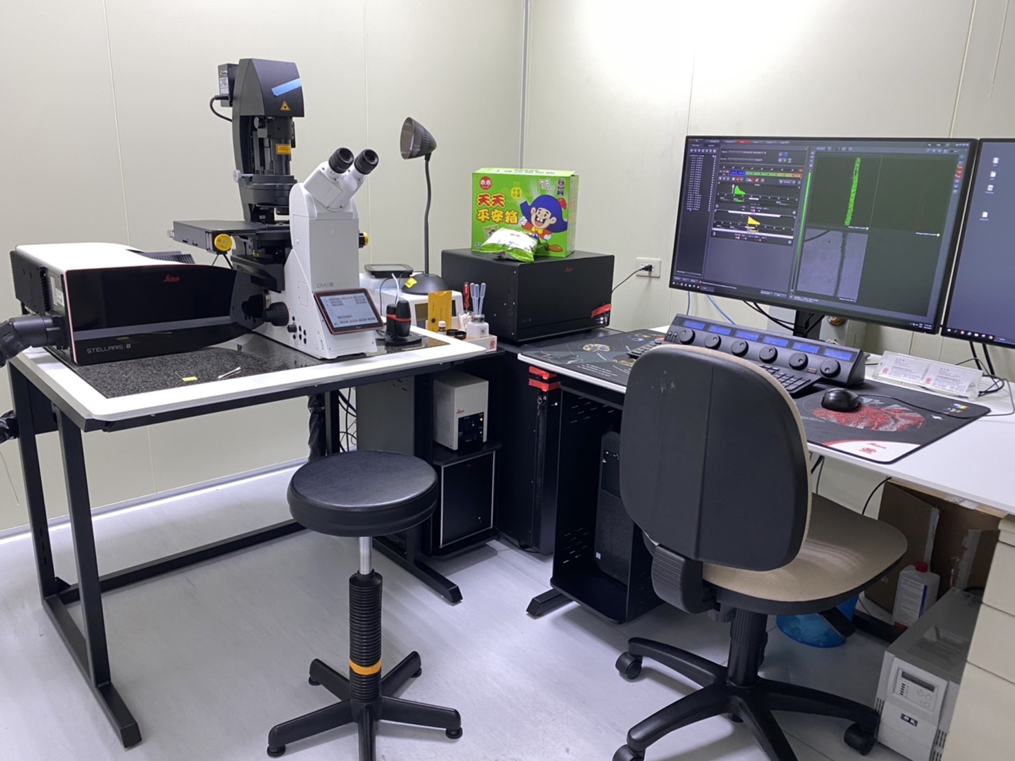 LEICA STELLARIS 8 (Invert, 2020), Diode 405nm; WHITE LIGHT LASER 440-790nm
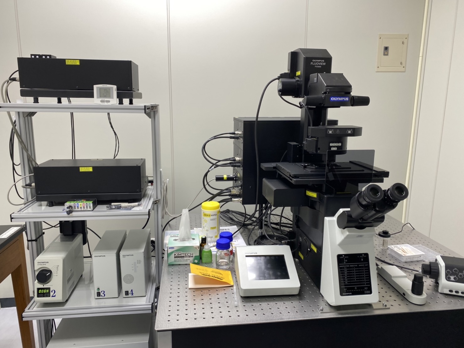 Olympus FV3000 (Invert 2022) lasers: 405 nm, 445 nm, 488 nm, 514 nm, 561 nm, 594 nm and 640nm