Spatial Tissue Imaging and Spatial Transcriptomics Division, Cell Biology Core Laboratory
Contact
- Chi-Chih Wu, Ph.D.
Committee
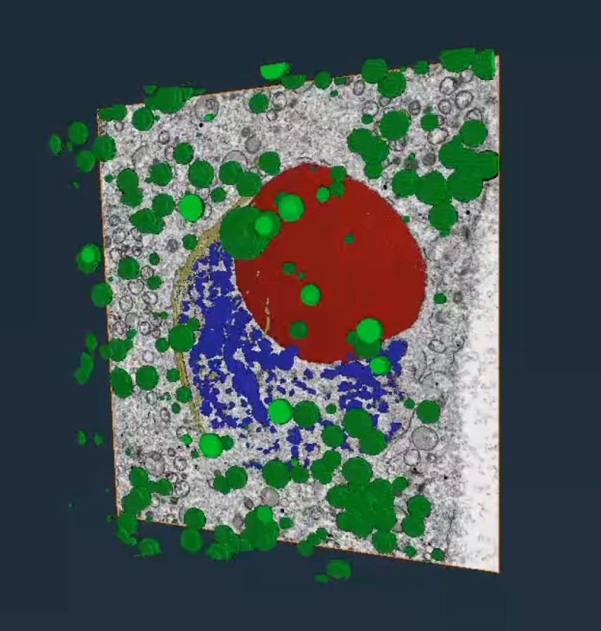
Three-dimensional reconstruction overlaid on an electron micrograph of a maize male meiocyte. Colors indicate the nucleolus (red), chromatin (blue), nuclear envelope (yellow), and vesicles (green).
Consultation and technical support for routine tissue sectioning, three-dimensional microscopy, and spatial transcriptomics.
Collaboration and Service Inquiries
Services are provided through project-based discussion and collaboration. Please contact us during experimental planning with your research objectives, sample types and sizes, and intended analyses so that we can assess technical feasibility and plan the next steps.
Services
1. Routine Histology Consultation and Support
We help assess tissue structures and sectioning requirements, select preparation methods suited to the samples and research objectives, and plan subsequent microscopy and analysis workflows.
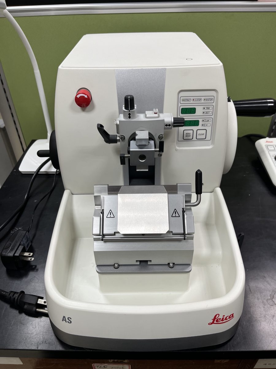
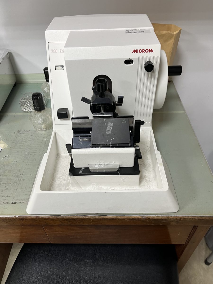
- Paraffin sectioning consultation: Consultation on tissue fixation, dehydration, paraffin embedding, and sectioning workflows to support tissue morphology studies and staining analyses.
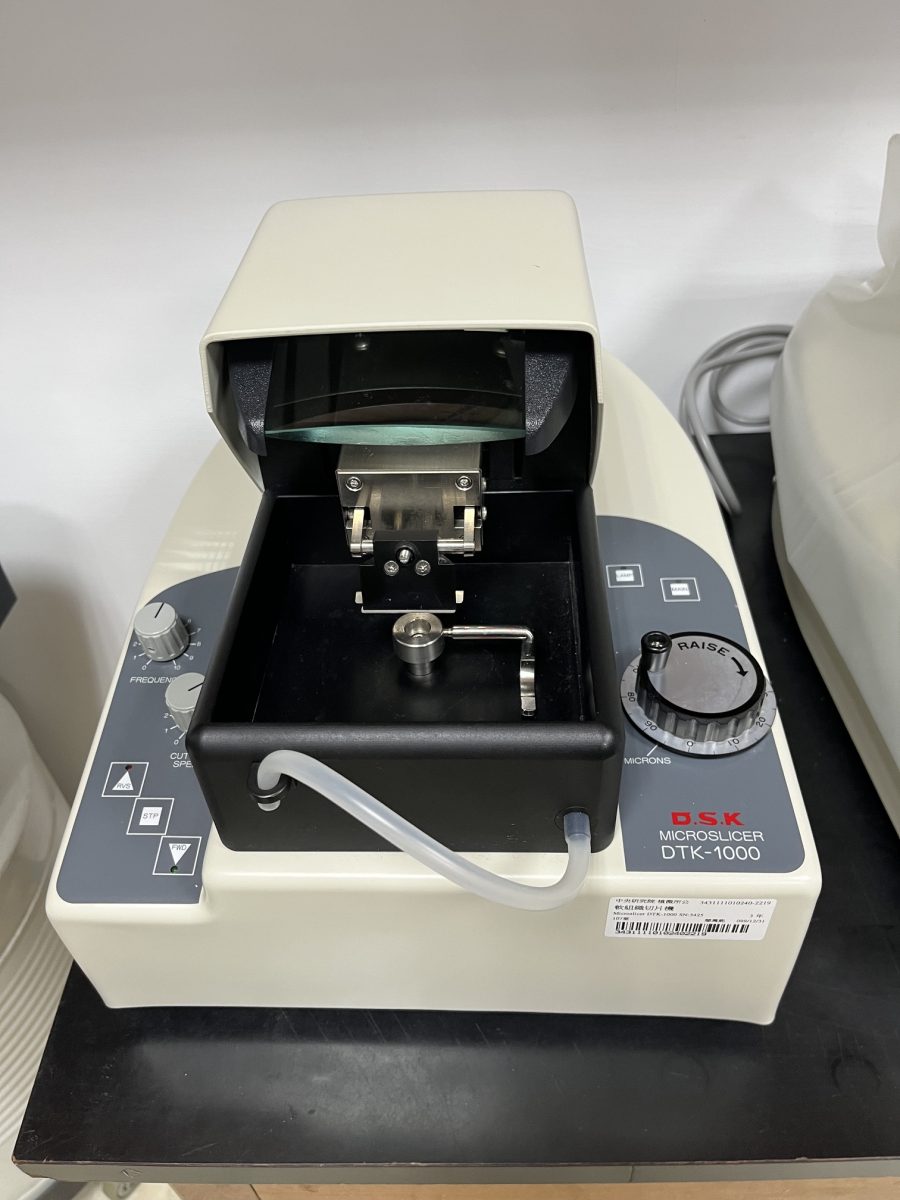
- Vibratome sectioning consultation: Consultation on sample handling, support embedding, and vibrating microtome settings to assess the preparation of thicker tissue sections and subsequent microscopy requirements.
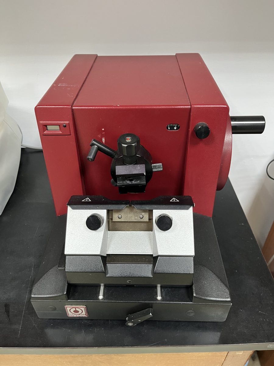
- Plastic sectioning consultation and services: Consultation and services for tissue fixation, dehydration, plastic embedding, and sectioning (1–10 µm thick) to support tissue morphology studies and staining analyses.
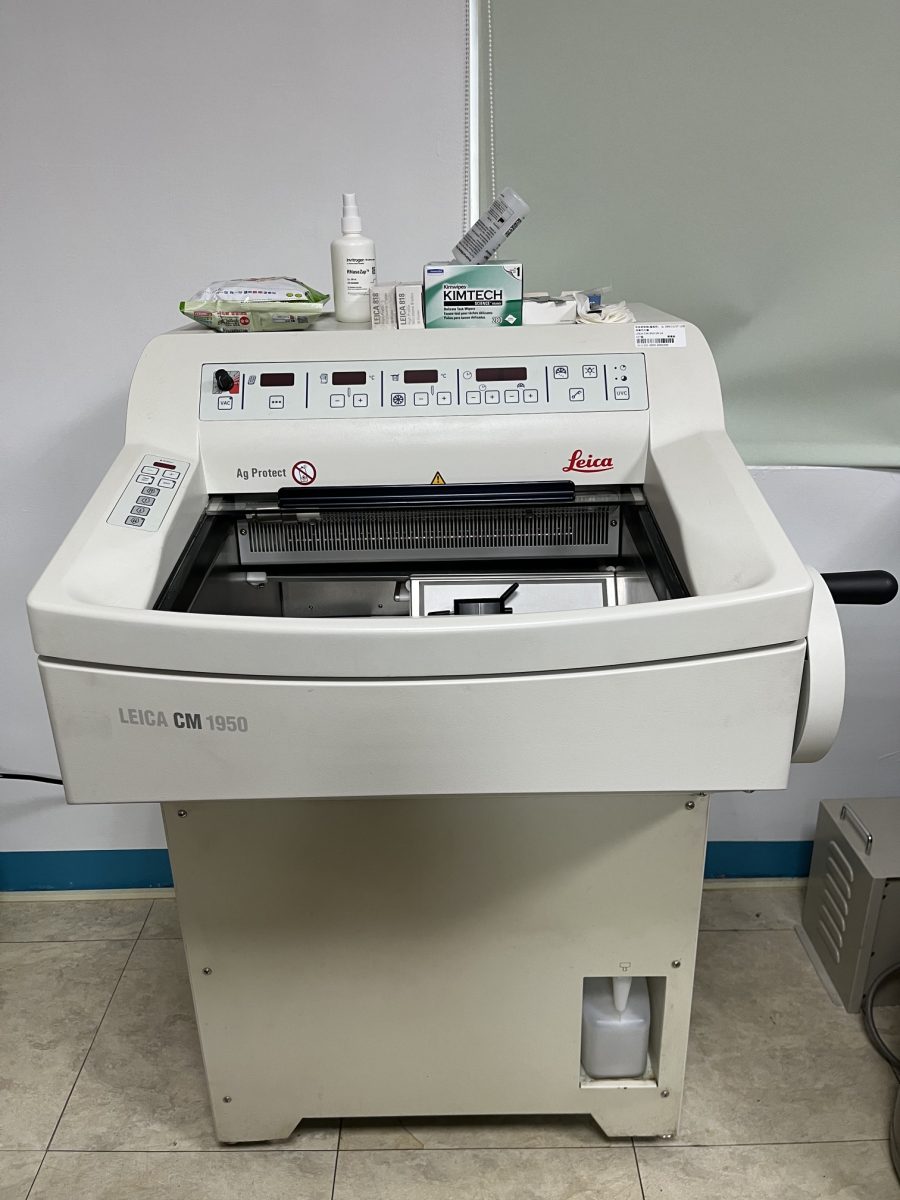
- Cryosectioning consultation and services: Technical consultation and cryosectioning services, including assessment of sample preparation, sectioning conditions, and downstream analysis requirements.
- Volume light microscopy: Consultation and technical support for volume light microscopy.
2. Volume Electron Microscopy
- Room-temperature volume EM: Sample preparation workflows for resin embedding and OTO staining are available, together with coordination of image acquisition requirements.
- Cryo-volume EM (small samples < 20 µm): Sample preparation workflows using plunge freezing and high-pressure freezing are available, with consultation on subsequent image acquisition and analysis.
- Cryo-volume EM (large samples > 20 µm): Sample preparation workflows using high-pressure freezing and cryo-FIB lift-out are available, with consultation on subsequent image acquisition and analysis.
Note — Focused Ion Beam Scanning Electron Microscopy (room-temperature and cryo-FIB-SEM) In collaboration with the Volume Electron Microscopy and Tomography Core Facility, we use the TF Hydra Bio platform to provide sample preparation under room-temperature and cryogenic conditions, volume electron microscopy, and related technical consultation. Services are arranged according to project requirements.
3. Cryo-Electron Tomography
We provide consultation and preparation services for Cryo-ET samples and cryo-lamellae. Workflows have been established for plunge-frozen samples and the high-pressure freezing grid-sandwich method. For large samples (> 20 µm), workflows for high-pressure freezing and cryo-FIB lift-out sample preparation have also been established. Cryo-lamella preparation and image acquisition are arranged at the Volume Electron Microscopy and Tomography Core Facility and the Cryo-Electron Microscopy Core Laboratory, with consultation on subsequent image acquisition and analysis.
We are also developing methods for subtomogram averaging and in situ molecular 3D reconstruction.
4. Spatial Transcriptomics
Combining anatomical assessment with spatial transcriptomics, we help research teams plan workflows from cryosectioning to sequencing library preparation and provide consultation and assistance with downstream data analysis. We currently primarily use the 10x Genomics Visium series of kits.
- Anatomical assessment of cryosections: Assessment of tissue structure and section integrity to help identify target regions for analysis.
- RNA integrity assessment: Assessment of RNA integrity in tissue sections in collaboration with the Genomics Technology Core Laboratory.
- mRNA capture and sequencing library preparation on Visium or Visium HD 3′ platforms: Conducted in collaboration with the Genomics Technology Core Laboratory.
5. In Situ Rolling Circle Amplification Labeling
Padlock probes and in situ rolling circle amplification labeling are used to analyze the distribution of target RNAs in tissue sections and quantify gene expression at cellular resolution.
- In situ labeling and expression analysis of target mRNAs: Assistance with planning in situ detection workflows for target genes and analyzing their expression across tissue regions and developmental stages.
- Quantification of gene expression at the cellular level: Consultation and technical support for in situ labeling, image acquisition, and signal quantification to assess target RNA expression at cellular resolution.
- Simultaneous detection of miRNA and mRNA: Assessment of the suitability of simultaneous miRNA and mRNA detection in tissues and assistance with planning expression analyses at the single-cell level.
Selected Publications
- Chi-Chih Wu, et al. Serial Cryosectioning for the Spatial Transcriptomics of Plant Tissues. Bio-protocol. (under review)
- Wye-Lup Kong, Chi-Chih Wu, Yi-Hua Chen, Jeng-Yi Li, Kai-Hsuan Ding, Yao-Ming Chang, Wen-Hsiung Li, and Mei-Yeh Jade Lu. Profiling maize embryonic leaf development and discovering new genes by high-resolution spatial long-read isoform sequencing. Nature Plants. 2026. https://doi.org/10.1038/s41477-026-02364-y
- Chi-Chih Wu, Ludvig Larsson, Kun-Ting Hsieh, Chun-Ping Yu, Yi-Hua Chen, Kai-Hsuan Ding, Ho-Chun Yang, Joakim Lundeberg, Chin-Min Kimmy Ho, Shu-Hsing Wu, Mei-Yeh Jade Lu, Wen-Hsiung Li. Serial spatial transcriptomes reveal regulatory transitions in maize leaf development. Plant Biotechnology Journal. 2025. https://doi.org/10.1111/pbi.70515
- Chi-Chih Wu, Kun-Ting Hsieh, Su-Ying Yeh, Yen-Ting Lu, Liang-Jwu Chen, Maurice S.B. Ku, and Wen-Hsiung Li. 2023. Simultaneous detection of miRNA and mRNA at the single-cell level in plant tissues. Plant Biotechnology Journal 21: 136–149. https://doi.org/10.1111/pbi.13931
- Chi-Chih Wu, Axel Klaesson, Julia Buskas, Petter Ranefall, Reza Mirazazdeh, Ola Söderberg, Jochen Wolf. 2019. In situ quantification of individual mRNA transcripts in melanocytes discloses gene regulation of relevance to speciation. Journal of Experimental Biology 222: jeb194431.
Instrument use and technical support are arranged according to project requirements. Please contact us in advance to confirm sample requirements and suitable workflows.
Instruments